Peptic Ulcer Disease (Gastric & Duodenal Ulcer / PUD)
An authoritative clinical and educational profile of Peptic Ulcer Disease (PUD), covering Helicobacter pylori infection, NSAID-induced mucosal injury, gastric and duodenal ulcer distinctions, constitutional homeopathic supportive management, and emergency red flags for gastrointestinal perforation, peritonitis, and massive upper GI hemorrhage.
Editorial review complete
Independent clinical validation is pending.
Quick Reference Facts
"Every gastric ulcer visualized on endoscopy must be biopsied and followed to complete endoscopic healing to rigorously exclude early gastric adenocarcinoma."
Visual guide
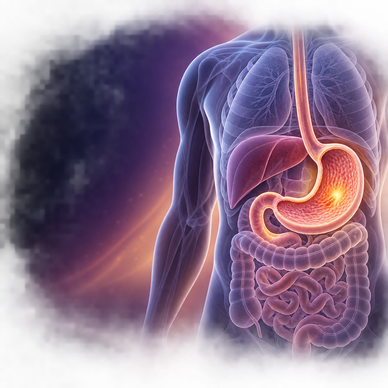
 Homeo Healthcare
 Homeo Healthcare
 Homeo Healthcare
In simple words
Peptic Ulcer Disease (PUD) refers to focal mucosal defects penetrating through the muscularis mucosae into the deeper layers of the stomach wall (gastric ulcer) or the first portion of the duodenum (duodenal ulcer). Primarily driven by Helicobacter pylori bacterial colonization or mucosal cyclooxygenase-1 (COX-1) inhibition by nonsteroidal anti-inflammatory drugs (NSAIDs), it manifests clinically as gnawing, burning epigastric pain, postprandial dyspepsia (indigestion and upper abdominal discomfort), early satiety, nausea, and vomiting.
A disruption in the integrity of the gastric or duodenal mucosa characterized by a circumscribed ulceration measuring ≥5 mm in diameter, extending into the submucosa or muscularis propria due to caustic acid-peptic digestion.
Common causes
- Helicobacter pylori infection (flagellated gram-negative microaerophilic bacterium producing urease, CagA/VacA cytotoxins, and chronic antral/corpus gastritis; accounts for ~70% of gastric ulcers and >85% of duodenal ulcers)
- Nonsteroidal Anti-inflammatory Drugs (NSAIDs, including aspirin, ibuprofen, naproxen, ketorolac, diclofenac; inhibit gastric mucosal prostaglandin synthesis via COX-1 suppression)
- Stress-related mucosal disease (severe physiological stress in ICU patients, sepsis, major trauma, extensive burns 'Curling ulcer', severe intracranial injury 'Cushing ulcer')
- Zollinger-Ellison syndrome (gastrinoma producing severe hypergastrinemia and refractory multiple atypical peptic ulcers)
Risk Factors
- Concurrent use of NSAIDs with systemic corticosteroids, anticoagulants, or antiplatelet agents (multiplies GI bleeding risk 4- to 12-fold)
- Tobacco smoking (inhibits pancreatic bicarbonate secretion, reduces mucosal blood flow, and accelerates ulcer recurrence)
- Chronic heavy alcohol consumption (direct mucosal barrier injury and stimulation of acid secretion)
- Advanced age (>65 years; higher prevalence of silent ulcers and severe complications)
- Prior history of documented peptic ulcer or upper gastrointestinal bleeding
Common symptoms
- Epigastric pain: rhythmic, sharp, gnawing, burning, or aching discomfort located between the xiphoid process and umbilicus
- Duodenal ulcer classic pattern: pain occurs 2 to 3 hours after meals or during the night (typically between 11 PM and 2 AM) and is characteristically relieved by food ingestion or antacids
- Gastric ulcer classic pattern: pain is often worsened or triggered soon after eating (within 15–30 minutes), leading to food avoidance and weight loss
- Postprandial bloating, abdominal distension, belching, early satiety, and nausea
- Occult blood in stool or iron deficiency anemia from chronic microscopic ulcer oozing
Clinical Red Flags
Seek urgent medical attention at an emergency department or primary care clinic if you present with any of the following symptoms:
- Acute Perforated Peptic Ulcer: sudden, catastrophic, explosive epigastric pain rapidly spreading across the entire abdomen, accompanied by rigid 'board-like' abdominal wall, rebound tenderness, and subdiaphragmatic free air on upright abdominal X-ray (surgical emergency requiring emergency laparoscopy/laparotomy)
- Massive Upper Gastrointestinal Hemorrhage: vomiting frank bright red blood or dark 'coffee-ground' emesis (hematemesis), or passing dark, tarry, sticky foul-smelling stools (melena), accompanied by tachycardia, dizziness, or hypovolemic shock (requires emergency endoscopic hemostasis and blood resuscitation)
- Gastric Outlet Obstruction (GOO): intractable projectile vomiting of undigested food eaten hours earlier, severe weight loss, and visible abdominal peristalsis due to chronic pyloric cicatricial scarring
Lifestyle & diet support
Strictly avoid NSAIDs and aspirin unless medically prescribed (substitute acetaminophen for simple pain relief where appropriate), eliminate cigarette smoking and tobacco use, abstain from heavy alcohol consumption, eat regular balanced meals rather than large heavy late-night meals, and practice stress-reduction techniques.
Treatment Approaches
Conventional Management
First-line medical therapy involves Proton Pump Inhibitors (PPIs: omeprazole, pantoprazole, esomeprazole; 4–8 weeks) to achieve gastric acid suppression. H. pylori eradication utilizes quadruple therapy (bismuth subcitrate + metronidazole + tetracycline + PPI for 14 days, or concomitant non-bismuth quadruple therapy). Discontinuation of offending NSAIDs or co-prescription of gastroprotective agents (PPIs, misoprostol). Endoscopic hemostasis (hemoclips, thermal coagulation, epinephrine injection) is indicated for actively bleeding ulcers.
Homeopathic Approach
Homeopathic constitutional and gastrointestinal remedies (such as Nux Vomica, Argentum Nitricum, Arsenicum Album, Hydrastis Canadensis, Phosphorus, Robinia Pseudacacia, Anacardium Orientale, Kali Bichromicum) serve as supportive care to soothe burning dyspepsia, ease nervous digestive irritability, and assist mucosal comfort alongside endoscopic evaluation and conventional H. pylori eradication therapy.
Frequently Asked Questions
Clinical & academic detailShow detailHide detail
Diagnosis & tests
Investigation Protocol
Esophagogastroduodenoscopy (EGD / upper endoscopy) is the definitive gold standard diagnostic investigation, allowing direct visualization of ulcer depth, size, and location, and mandatory multiple biopsies of gastric ulcers to exclude gastric adenocarcinoma or lymphoma. H. pylori testing includes urea breath test (UBT), stool antigen test (HpSA), or endoscopic mucosal biopsy with rapid urease testing (RUT / CLO test).
Differential Diagnosis
Differentiate Peptic Ulcer Disease from Functional Dyspepsia, Gastroesophageal Reflux Disease (GERD), Biliary Colic / Acute Cholecystitis, Acute or Chronic Pancreatitis, Gastric Malignancy, Mesenteric Ischemia, and Coronary Artery Disease (inferior wall myocardial infarction presenting as epigastric burning).
Differential Diagnosis Matrix
Differentiate Peptic Ulcer Disease from Functional Dyspepsia, Gastroesophageal Reflux Disease (GERD), Biliary Colic / Acute Cholecystitis, Acute or Chronic Pancreatitis, Gastric Malignancy, Mesenteric Ischemia, and Coronary Artery Disease (inferior wall myocardial infarction presenting as epigastric burning).
Reference Citations & Evidence Sources
Classical Homeopathic Literature
- CIT-0007Hahnemann S.. "The Chronic Diseases: Their Peculiar Nature and Their Homoeopathic Cure." Adolph Arnold (1828).
Materia Medica & Keynotes
- CIT-0004Hahnemann S.. "Materia Medica Pura." Adolph Arnold (1811).
- CIT-0005Kent J. T.. "Lectures on Homoeopathic Materia Medica." Boericke & Tafel (1905).
- CIT-0006Boericke W.. "Pocket Manual of Homoeopathic Materia Medica." Boericke & Runyon (1901).
Clinical Reviews & Textbooks
- CIT-0023National Center for Complementary and Integrative Health. "Homeopathy: What You Need To Know." National Institutes of Health (2021).
No clinical connections registered for this topic.
Seeking Expert Consultation?
Book an online video session with Dr. Narayan Jethwani to get an individualized constitutional homeopathic protocol tailored to your case.
Medical Safety Disclaimer
All content on the Homeo Healthcare platform is strictly for educational purposes and is not personal medical advice, diagnosis, or treatment. Homeopathic remedy considerations are provided for clinician review or require individualized consultation with a qualified physician. Never delay seeking professional medical advice or emergency medical care due to content you have read on this website.